
4.16
Surgical Wound Dehiscence
Obstetrics & Gynaecology Protocols, Version 3.0, 2017. Chapter 4, Gynaecology.
Clinical description
Dehiscence occurs when fascia, subcutaneous tissue and skin separate prior to healing. Risk factors include: haematoma, seroma, excessive intra-abdominal pressure (i.e. coughing or vomiting), DM, malignancies, anaemia, infection, immunosuppression, poor technique and inappropriate suture. Haematomas and seromas predispose to infection as they can cause the incision to separate and allow bacteria to gain access to deeper layers.
Steps to prevent surgical wound complications
- Maintain haemostasis
- Handle tissues gently
- Remove devitalized tissue
- Use monofilament suture, taking bites with at least 1 cm of tissue
- If subcutaneous tissue ≥ 2 cm depth, then close dead space with subcutaneous suture in Camper’s fascia
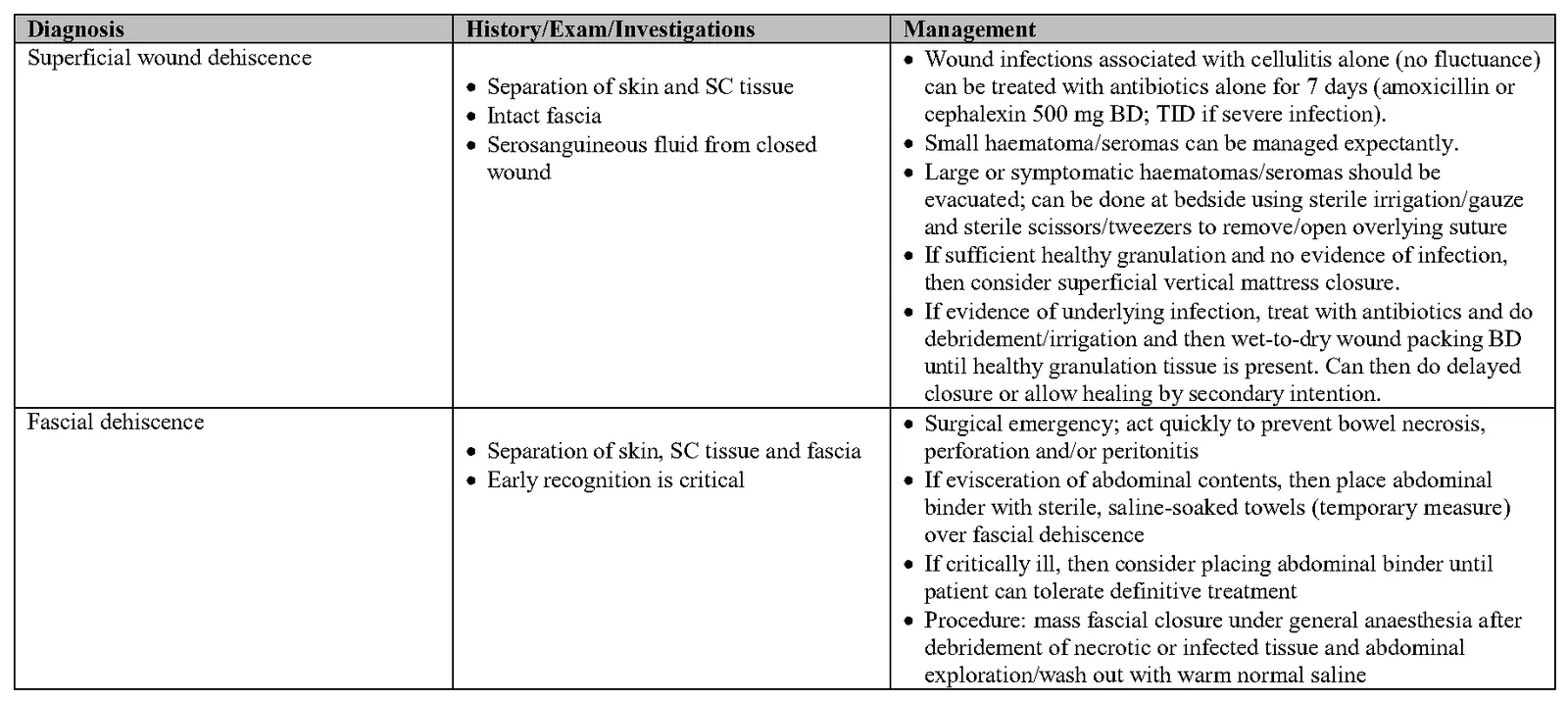
Diagnosis History/Exam/Investigations Management Superficial wound dehiscence • Wound infections associated with cellulitis alone (no fluctuance)
- Separation of skin and SC tissue can be treated with antibiotics alone for 7 days (amoxicillin or
- Intact fascia cephalexin 500 mg BD; TID if severe infection).
- Serosanguineous fluid from closed • Small haematoma/seromas can be managed expectantly.
wound • Large or symptomatic haematomas/seromas should be evacuated; can be done at bedside using sterile irrigation/gauze and sterile scissors/tweezers to remove/open overlying suture
- If sufficient healthy granulation and no evidence of infection,
then consider superficial vertical mattress closure.
- If evidence of underlying infection, treat with antibiotics and do
debridement/irrigation and then wet-to-dry wound packing BD until healthy granulation tissue is present. Can then do delayed closure or allow healing by secondary intention.
Fascial dehiscence • Surgical emergency; act quickly to prevent bowel necrosis,
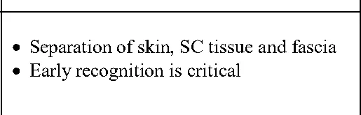
- Separation of skin, SC tissue and fascia perforation and/or peritonitis
- Early recognition is critical • If evisceration of abdominal contents, then place abdominal
binder with sterile, saline-soaked towels (temporary measure) over fascial dehiscence
- If critically ill, then consider placing abdominal binder until
patient can tolerate definitive treatment
- Procedure: mass fascial closure under general anaesthesia after
debridement of necrotic or infected tissue and abdominal exploration/wash out with warm normal saline
Tables and figures
Check doses against the printed guideline and your clinical judgement before treating a patient. Spotted an error? Report it from the contact links below.
Need this without data?
The app holds every guideline on your device, with calculators, bookmarks and notes.