
1.4
Gestational Trophoblastic Disease
Obstetrics & Gynaecology Protocols, Version 3.0, 2017. Chapter 1, Early Pregnancy Complications.
Clinical description
Gestational trophoblastic disease (GTD) are diseases that arise from abnormal proliferation of placental trophoblastic cells. GTD includes hydatidiform mole (complete or partial) andGestational Trophoblastic Neoplasia (GTN), choriocarcinoma, and placental site trophoblastic tumours. Following molars pregnancy about 20% of women will develop malignant disease (GTN). GTN is more common in women with complete molar pregnancies, large theca-luetin cysts (>5cm), extremely enlarged uterus, >40 years old, very high hCG levels and/or prior history of GTD.
Molar Pregnancy:
Signs and symptoms
Signs/Symptoms: 1st trimester bleeding, uterine size/date discrepancy, sudden increase in uterine size, hyperemesis, passage of vesicles, early gestational hypertension/pre-eclampsia, thyrotoxicosis and greatly elevated hCG.Classically shows "snowstorm" pattern. on ultrasound scan.
History/Exam/Investigations:Patients are at risk of hyperthyroidism/thyroid storm, anemia, coagulopathies, pre- eclampsia/eclampsia. Ask appropriate questions to evaluate for these conditions. Obtain complete vital signs, evaluate uterine size, cervical dilatation and evidence of active bleeding or passage of tissue. Obtain ultrasound scan if not already done. Very high hCG can cause a urine pregnancy test can be falsely negative in molar pregnancies- consider dilution of the urine and re-testing if clinically suspicion is high.
Treatment
- Order: group and cross-match, FBC, clotting tests, chest x-ray (to rule out lung metastasis) and quantitative
hCG if available.
- Ideally, management should take place in a facility where blood transfusion and ICU care are available as
hemorrhage and respiratory distress are possible complications.
- If asymptomatic, then schedule suction D&C under ultrasound-guidance (if available) with anesthesia with
oxytocin infusion and misoprostol ready due tohigh risk of haemorrhage.
- If actively aborting, send patient to theatre immediately and do D&C under ultrasound-guidance (if
available), with oxytocin and misoprostol ready.
- Suction D&C under ultrasound-guidance with the largest suction canula possible is the preferred method of
evacuation. Sharp curettage ALONE should NOT be performed.
- Risk of hemorrhage and uterine perforation are high.
- Oxytocin infusion should be started after dilation of cervix and continued for several hours
postoperatively.
- Hysterectomy can be considered in cases where childbearing is complete, but does not negate the need for
close follow-up.
- Give Antibiotics (Doxycycline 100 mg BD x 3 days or Metronidazole 400 mg BD x 5 days) for
prophylaxis
- Send tissue for pathologic examination.
Follow-up
- Follow up all cases for serial evaluation.
- Stress importance of avoiding pregnancy until follow-up is complete and encourage use of a
highly effective form of birth control (avoid IUCD until hCG is undetectable).
- Review histology with patient at 1st follow-up visit.
- At each visit:
Send urine for pregnancy test (should be negative by 60-100 days post evacuation)
- Follow-up serum Hcg instead of UPT if available.
Conduct bimanual pelvic exam to assess uterine size Conduct speculum exam of vagina and suburethral area for metastases. If noted, do not biopsy as this can lead to hemorrhage.
Conduct ultrasound scan to evaluate for intrauterine and extrauterine signs of disease.
- Monthly follow-up until 1 year after evacuation.
Clinical description
- Presumed diagnosis of persistent gestational trophoblastic neoplasm if UPT or Hcg remains
positive 4 months after evaluation or if there is evidence of molar re-occurrence or metastatic lesions on imaging. (Make sure to rule out new pregnancy before initiation of treatment).
- In subsequent pregnancies, counsel on the importance of early antenatal care, order early US to
look for recurrent mole (10 times more common in patients with prior molar pregnancy).
Persistant Gestational Trophoblastic Neoplasm: The vast majority of women with persistent GTN can be cured with chemotherapy +/- surgery and radiation therapy. Timely diagnosis and appropriate treatment is key.
Remember, although GTN is most common following a molar pregnancy, it can follow any pregnancy event. Suspect GTN in any woman who has bleeding >6 weeks after pregnancy or who has metastatic disease with no known primary and a positive UPT with no intrauterine pregnancy.
Initial Investigations: vaginal ultrasound, chest X-ray, vaginal examination.
If metastatic lesions are found on Pelvic USS, CXR or vaginal exam, perform liver and brain imaging if possible.
Treatment
- All patients with persistent disease should be referred to medical oncology for initiation of chemotherapy.
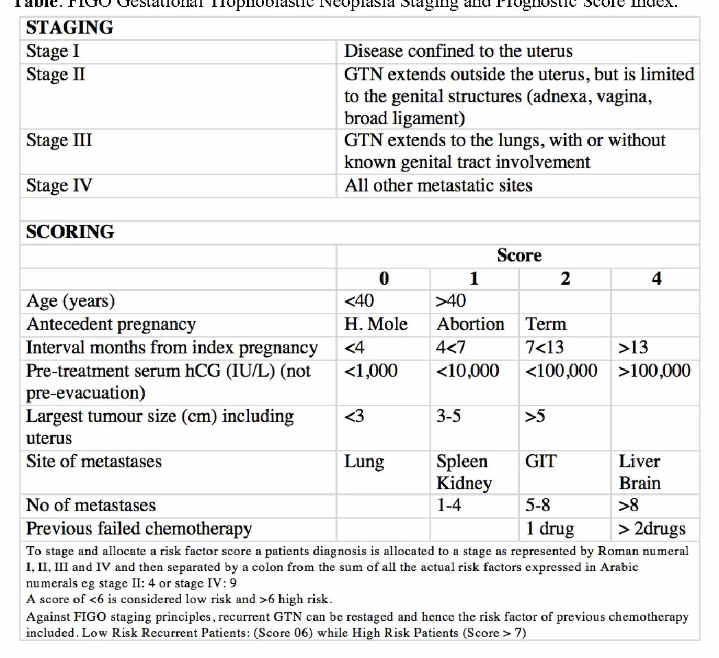
The chemotherapy regimen should be based on risk assessment using the FIGO Prognostic Score Index.
Score of ≤6 is deemed low-risk and needs single-agent chemotherapy (typically methotrexate). A score >6 is high-risk and should have initial multiple drug chemotherapy.
- Hysterectomy can be considered in women who have completed child bearing and have disease confined to
the uterus. It is required in patients with Placental Site Trophoblastic Tumor which is less responsive to chemotherapy. Hysterectomy is not effective against metastatic disease.
- Radiation therapy may be necessary for patients with brain metastasis.
Table: FIGO Gestational Trophoblastic Neoplasia Staging and Prognostic Score Index.
Tables and figures
Check doses against the printed guideline and your clinical judgement before treating a patient. Spotted an error? Report it from the contact links below.
Need this without data?
The app holds every guideline on your device, with calculators, bookmarks and notes.