
1.2
Cervical Insufficiency
Obstetrics & Gynaecology Protocols, Version 3.0, 2017. Chapter 1, Early Pregnancy Complications.
Clinical description
Cervical insufficiency is a clinical diagnosis characterized by painless cervical dilatation and spontaneous mid trimester pregnancy loss (14 weeks to 24 weeks gestation), in absence of labour and other causes.
Risk factors for cervical insufficiency
- History- past history of recurrent midtrimester losses, cervical surgery (cone biopsy, large
loop excision of the transformation zone, D&C
- Structural- congenital uterine abnormalities- septate, bicornuate uterus
- Physical – cervical shortening <25mm before 28 weeks on transvaginal US, cervical tears on
physical examination Indicationsfor cervical cerclage History-indicated cervical cerclage
- 3 or more spontaneous consecutive midtrimester pregnancy losses that have a typical history of
cervical insufficiency
- Placement of history-indicated cerclage is ideally at 13-14weeks gestation
Ultrasound-indicated cervical cerclage
- This is offered to women with previous preterm birth after assessment through cervical length
screening
- Cervical length screening- serial transvaginal cervical length assessment between 14-24 weeks
gestation. Cervical cerclage is offered before 24 weeks gestation in those women found with a short cervix <25mm.
Physical examination-indicated cervical cerclage
- Physical examination-indicated cerclage is the placement of cervical cerclage in women in the second
trimester who present with cervical dilatation in absence of labour or placental abruption., Contraindications to cervical cerclage insertion
- Labour, vaginal bleeding, ruptured membranes, chorioamnionitis, placental abruption
- Lethal fetal anomalies, multiple pregnancy
- Cervical dilatation >4cm
Investigations
History
- Thorough history – obstetric, medical, surgical, social
- Exclude other causes
- Medical: uncontrolled diabetes, hypertension, thyroid
- Fetal anomaly, infection, abruption
Exam
- Thorough physical and obstetric evaluation
- Vaginal speculum: scarring, tears, cervical length, infection, cervical dilatation, exclude rupture of
membranes
- Wet mount/high vaginal swab: exclude infections
- Urine dipstick
Investigations
- Antenatal screening tests: syphilis, HIV, Rhesus
- Transvaginal US: for cervical length measurement
- Abdominal US: for viability, dating pregnancy, rule out multiple gestation and lethal congenital
anomalies Timing of cervical cerclage insertion
- Between 13-24 weeks
- History-indicated cerclage should be placed as early as possible, starting at 13 weeks
Follow-up
- On complications: preterm prelabour rupture of membranes, pregnancy loss, bleeding, infection,
hospitalization, procedural risks Preoperative and postoperative management
- Discuss with Consultant about whether to give Erythromycin 500 mg or Clindamycin 600 mg with
Indomethacin 100 mg PO or PR q12 hours for 24 hours post-operative
- If this regimen is given, patient should be kept in hospital while receiving treatment
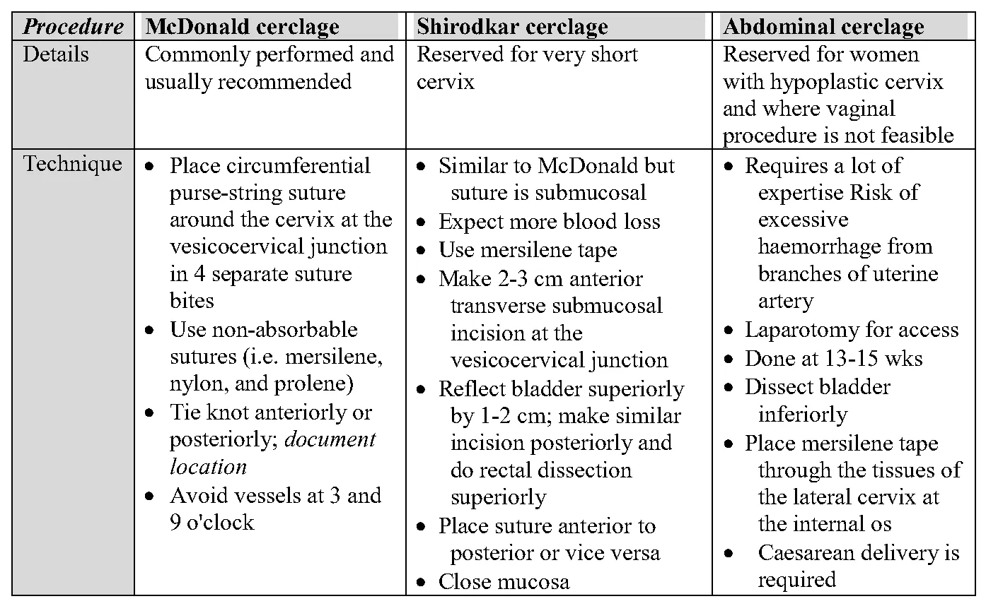
Surgical procedure There are three main techniques used for cervical cerclage insertion described below. The most commonly- used and easiest to learn is the McDonald cerclage.
Procedure McDonald cerclage Shirodkar cerclage Abdominal cerclage Details Commonly performed and Reserved for very short Reserved for women usually recommended cervix with hypoplastic cervix and where vaginal procedure is not feasible Technique • Place circumferential • Similar to McDonald but • Requires a lot of purse-string suture suture is submucosal expertise Risk of around the cervix at the • Expect more blood loss excessive vesicocervical junction • Use mersilene tape haemorrhage from in 4 separate suture • Make 2-3 cm anterior branches of uterine bites transverse submucosal artery
- Use non-absorbable incision at the • Laparotomy for access
sutures (i.e. mersilene, vesicocervical junction • Done at 13-15 wks nylon, and prolene) • Reflect bladder superiorly • Dissect bladder
- Tie knot anteriorly or by 1-2 cm; make similar inferiorly
posteriorly; document incision posteriorly and • Place mersilene tape location do rectal dissection through the tissues of
- Avoid vessels at 3 and superiorly the lateral cervix at
9 o'clock • Place suture anterior to the internal os posterior or vice versa • Caesarean delivery is
- Close mucosa required
Post-operative management
- Immediate post-operative care includes
- Analgesia
- Antibiotic continuation
- Normal diet unless complication
- Discharge next day after assessment
- Bedrest- where clinically indicated but not as a routine for all patients
- Routine ANC unless otherwise indicated with repeated counseling regarding infection, labour, PROM
Antenatal care
- Monthly
- Preterm labour: tocolysis for steroid administration if no contraindications and <34 weeks
- After 34 weeks, remove cervical cerclage and do not give tocolysis
- PPROM: remove cervical cerclage
Cervical cerclage removal
Clinical description
- If no problems, routine removal at 36-37 weeks gestation
- Other indications:
- Regular contractions -> then follow preterm labour protocol
- PPROM ->then follow PPROM protocol
- Intrauterine fetal death -> then follow IUFD protocol
- Antepartum hemorrhage
- Chorioamnionitis
Tables and figures
Check doses against the printed guideline and your clinical judgement before treating a patient. Spotted an error? Report it from the contact links below.
Need this without data?
The app holds every guideline on your device, with calculators, bookmarks and notes.